Ultra-short X-ray flashes explore the nano world
Characterization of extremely short X-ray pulses paves the road to new applications
Ultra-short and extremely strong X-ray flashes, as produced by free-electron lasers, are opening the door to a hitherto unknown world. Scientists are using these flashes to take “snapshots” of the geometry of tiniest structures, for example the arrangement of atoms in molecules. To improve the spatial and time resolution further requires knowledge about the precise duration and intensity of the X-ray flashes.
An international team of scientists has now tackled this challenge. Using the Linac Coherent Light Source (LCLS) in Stanford, California, scientists from the Technische Universität München (TUM), the Max Planck Institute of Quantum Optics (MPQ), and from the Deutsches Elektronen Synchrotron (DESY) succeeded in measuring the duration of ultrashort X-ray pulses (Nature Photonics, DOI: 10.1038/NPHOTON.2014.278, 24 November 2014).
X-ray flashes are a unique scientific tool. They are generated by accelerating electrons to very high energy levels in kilometer-long vacuum tubes, so-called linear accelerators, and then deflecting them with specially arranged magnets. In the process the particles emit X-ray radiation that amplifies itself until an ultra-short and intensive X-ray flash is released. Researchers use these X-ray flashes to resolve structures as small as one ten billionth of a meter (0.1 nanometer) in size. That is roughly the diameter of a hydrogen atom. In this way, biomolecules, for example, can be imaged at extremely high resolution, providing new insight into the nano cosmos of nature.
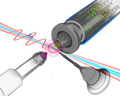
 the scientists investigate the temporal structure of the X-ray pulse (pictured in blue). When the X-ray flash hits a gas atom it knocks electrons out of the innermost shell, setting them free. The electrons are then accelerated or decelerated by the electrical field of the infrared light pulse. As the energies of the electrons (green) vary depending on the time of their emission the duration of the X-ray pulse that has triggered the reaction can be deduced.")
Using two quickly sequenced flashes the researchers can even obtain information on structural changes during reactions. The first laser flash triggers a reaction while the second measures structural changes during the reaction. For this it is essential to know the precise duration and temporal intensity progression of the X-ray flashes. However, hitherto it has not been possible to measure the ultra-short pulses directly.
Researchers at the Technische Universität München (TUM), the Hamburg Center for Free-Electron Laser Science (CFEL) and the Max Planck Institute of Quantum Optics (MPQ) in Garching, in collaboration with other colleagues, have now developed just such a methodology. The respective experiments were done at the SLAC National Accelerator Laboratory in California (USA) by a team headed by Professor Reinhard Kienberger, Dr. Wolfram Helml (TUM) and Dr. Andreas Maier (CFEL). The CFEL is a collaboration facility of the German Electron Synchrotron (DESY), the University of Hamburg and the Max Planck Society.
The scientists determined the duration of the X-ray flashes by modifying a process originally developed to measure ultra-short flashes of light. The physicists directed the X-ray flashes into a vacuum chamber filled with a few atoms of an inert gas. There they superimposed the flashes with 2.4 micrometer wavelength pulses of infrared light. When the X-ray flashes hit a gas atom they knock electrons out of the innermost shell, setting them free. The electrons are then accelerated or decelerated by the electrical field of the infrared light pulse. The change in an electron’s velocity is a function of when the light intercepts the electron and the electrical field strength at the moment of interception.
Since electrons are set free during the full duration of an X-ray flash, electrons emitted at different points in time “feel” different field strengths of the periodically oscillating infrared light. As a result they are accelerated at varying rates. The physicists can then calculate the duration of the original X-ray flash from the different arrival times of the electrons in a detector.
Using this approach, the researchers were able to determine the average upper limit of the pulse duration of their X-ray flashes at around 4.4 femtoseconds – a femtosecond is a millionth of a billionth of a second (10-15 seconds). In addition, the researchers obtained insight into the structure of the X-ray flashes. Characteristic for the strong X-ray pulses in free-electron lasers is their randomly changing pulse form. A typical X-ray pulse comprises multiple contiguous shorter “X-ray spikes.” The number and intensity of these spikes varies from one shot to the next. For the first time ever, the researchers managed to measure these ultra-short sub-peaks directly and confirm predictions that the individual flashes last only around 800 attoseconds – an attosecond is a billionth of a billionth of a second (10-18 seconds). The new methodology makes possible the detailed, direct measurement of X-ray pulses and augments methodologies for determining pulse shape and length indirectly from the structure of the electron packets used to generate the flashes.
The enhanced X-ray pulse measurement technology may also find application at the new Center for Advanced Laser Applications (CALA) at the Garching campus. Researchers there are working on, among other things, generating even shorter X-ray pulses using high-energy lasers. Pulses with a duration of only a few attoseconds, would allow researchers to take “snapshots” of even faster processes in nature, like the motion of electrons around atomic nuclei.
However, X-ray flashes provide not only basic research with new perspectives. Medicine could also profit from the technology. “Ultra-short laser-like X-ray pluses serve not only the investigation of the fastest physical processes in the core of matter, but could, because of their extremely high intensity, also be used to destroy tumors following X-ray diagnosis,” explains Reinhard Kienberger, professor for laser and X-ray physics at TU München and leader of the research consortium.
Thorsten Naeser


